
Abstract
Introduction
To evaluate the impact of a basic point-of-care ultrasound (POCUS) course on physicians’ clinical use of POCUS in routine medical practice.
Methods
This retrospective cross-sectional survey included physicians who attended one of 48 basic POCUS courses organized by the Emergency Medicine Association of Türkiye between 2019 and 2023. Among 610 physicians with available contact information, 201 completed the electronic survey, resulting in a response rate of 33%. The survey assessed participants’ demographics, previous ultrasound training, and self-reported changes in POCUS usage before and after the course.
Results
The mean age of participants was 30.9±4.6 years, and 83.6% were emergency medicine specialists. A statistically significant increase in POCUS use was reported across all modalities following the course (p<0.001). The most prominent increases occurred in hepatobiliary (+58.8%), lower extremity deep vein thrombosis (+54.0%), and abdominal aorta (+50.8%) examinations. Use of POCUS for interventional procedures also rose substantially, particularly for lumbar puncture (+81.8%) and peripheral nerve blocks (+66.6%). Participants also reported enhanced diagnostic confidence, procedural competence, and integration of POCUS into decision-making.
Conclusion
A short, structured POCUS course led to a meaningful increase in the clinical use of ultrasound across various diagnostic and interventional domains. Despite limitations such as recall bias and lack of follow-up assessment, the findings highlight the potential of focused training to improve practice patterns. Future course designs may benefit from incorporating certification components, objective structured clinical examination-style evaluations, and opportunities for supervised practice. Follow-up or refresher training may further support skill retention and long-term integration.
Keywords: Point-of-care systems, ultrasonography, medical education, emergency medicine, clinical competence, surveys and questionnaires
Introduction
Point-of-care ultrasound (POCUS) is a bedside diagnostic method increasingly integrated into clinical practice, especially in emergency medicine. It enables rapid assessment in various clinical scenarios, including chest pain, dyspnea, abdominal pain, hypotension, and during interventional procedures such as central line placement and thoracentesis (-). However, as an operator-dependent modality, its accuracy hinges on the user’s technical and interpretative competence.
While POCUS has been incorporated into emergency medicine residency training in many countries, standardized training curricula for other specialties remain limited and heterogeneous (-). Structured educational programs based on established guidelines have demonstrated improvements in diagnostic performance and user confidence (-). However, there remains a gap in the literature regarding the long-term impact of short-duration POCUS courses, particularly in non-standardized training settings ().
This study aims to evaluate the effect of a basic POCUS course on the clinical ultrasound usage patterns of physicians. The primary objective is to measure the change in POCUS use after the course, while the secondary objective is to identify which ultrasound applications were most frequently adopted into practice.
Methods
Study Design and Population
This study was designed as a retrospective cross-sectional survey. The target population consisted of 1,324 physicians who had participated in one of 48 basic POCUS courses organized by the Ultrasound Section of the Emergency Medicine Association of Türkiye between 2019 and 2023. Despite missing or outdated contact information, 610 physicians could be reached, and 201 of them completed the survey (response rate: 33%). To evaluate the sustained impact of the training, the survey was administered at least six months after participants had completed the POCUS course.
Survey Instrument
A 28-item electronic survey was used to collect data. It included questions on demographic characteristics, specialty, years of experience, previous ultrasound training, and availability of ultrasound devices in the workplace. Physicians retrospectively evaluated their POCUS use before and after the course, including frequency and indication of use. The survey included both multiple-choice and 5-point Likert scale items, assessing perceived changes in diagnostic confidence, procedural competence, and integration of POCUS into clinical decision-making.
POCUS Course Content
The basic POCUS course consisted of a combination of theoretical and practical sessions, delivered over a two-day format. The curriculum included ultrasound physics and knobology, Extended Focused Assessment with Sonography for Trauma (E-FAST), hepatobiliary, urinary, and genital systems, abdominal aorta, inferior vena cava, echocardiography, lower extremity deep vein thrombosis (DVT), and ultrasound-guided procedures such as vascular access and lumbar puncture. Hands-on training was conducted under supervision, with participants practicing image acquisition and interpretation on live standardized patients. Interventional techniques were practiced on gelatin-based handmade phantom models to simulate vascular and lumbar procedures.
Statistical Analysis
Descriptive statistics were used to summarize participant characteristics. Continuous variables were expressed as mean ± standard deviation and categorical variables as frequencies and percentages. The normality of continuous variables was assessed using the Shapiro-Wilk test. McNemar and McNemar-Bowker tests were used to compare pre- and post-course changes in paired categorical variables. A p-value of <0.05 was considered statistically significant. All statistical analyses were conducted using SPSS version 25.0 (IBM Corp., Armonk, NY, USA).
Ethical Considerations
The study was approved by the University of Health Sciences Türkiye, Fatih Sultan Mehmet Training and Research Hospital Clinical Research Ethics Committee (approval number: 2023/19, date: 12.10.2023), and conducted in accordance with the principles of the Declaration of Helsinki (2013 revision). Informed consent was obtained from all participants prior to their inclusion in the study.
Results
The mean age of the participants was 30.9±4.6 years, and 53.7% were male. Most of the respondents were emergency medicine specialists (83.6%), followed by internal medicine physicians (6.5%) and general practitioners (3.5%). The average professional experience was 6.2±4.4 years. A total of 47.8% of the participants were working in training and research hospitals, 31.8% in university hospitals, and 14.9% in state hospitals.
Among the participants, 54.2% reported receiving ultrasound education for the first time, and 93% had access to an ultrasound device at their institutions. The most commonly available probe types were curvilinear (35.9%), linear (35.2%), and sector (27.8%) ().
Table 1. Demographic characteristics of physicians participated in the study
Characteristic | Value |
|---|---|
Age (years), mean ± SD (range) | 30.9±4.6 (25-55) |
Gender, n (%) | - |
Female | 93 (46.3) |
Male | 108 (53.7) |
Medical specialty, n (%) | - |
Emergency medicine | 168 (83.6) |
Internal medicine | 13 (6.5) |
General practice | 7 (3.5) |
General surgery | 4 (2.0) |
Pediatrics | 4 (2.0) |
Anesthesia and reanimation | 3 (1.5) |
Neurology | 1 (0.5) |
Other | 1 (0.5) |
Years in practice, mean ± SD | 6.2±4.4 |
Institution type, n (%) | - |
Training and research hospital | 96 (47.8) |
University hospital | 64 (31.8) |
State hospital | 30 (14.9) |
City hospital | 6 (3.0) |
Private hospital | 5 (2.5) |
Prior ultrasound training, n (%) | - |
Yes | 92 (45.8) |
No | 109 (54.2) |
Training year, n (%) | - |
2019 | 27 (13.4) |
2020 | 6 (3.0) |
2021 | 22 (10.9) |
2022 | 72 (35.8) |
2023 | 74 (36.8) |
USG device availability, n (%) | - |
Yes | 187 (93.0) |
No | 14 (7.0) |
Available USG probes, n (%) | - |
Curvilinear (convex) | 190 (35.9) |
Linear | 186 (35.2) |
Sector (cardiac) | 147 (27.8) |
Other | 6 (1.2) |
The results are expressed in terms of mean ± standard deviation (minimum and maximum) and n (%) values
SD: Standard deviation, USG: Ultrasonography
As shown in , participants reported statistically significant increases in their self-reported use of POCUS, along with improvements in diagnostic confidence, equipment familiarity, and patient management after the course (p<0.001).
Table 2. Physicians views on their POCUS use before and after the basic POCUS course
Parameters | Before the course | After the course | p value* |
|---|---|---|---|
I use POCUS in my patient management when indicated | |||
Never | 44 (21.9) | 1 (0.5) | <0.001 |
Seldomly | 93 (46.3) | 8 (4) | |
Occasionally | 42 (20.9) | 40 (19.9) | |
Frequently | 16 (8) | 112 (55.7) | |
Always | 6 (3) | 40 (19.9) | |
Incorporating POCUS into my patient management improves my diagnostic skills | |||
I strongly disagree | 3 (1.5) | 1 (0.5) | <0.001 |
I disagree | 14 (7) | 0 | |
I neither agree nor disagree | 78 (38.8) | 1 (0.5) | |
I agree | 75 (37.3) | 62 (30.8) | |
I strongly agree | 31 (15.4) | 137 (68.2) | |
I am familiar with the USG device and its technical features | |||
I strongly disagree | 68 (33.8) | 1 (0.5) | <0.001 |
I disagree | 83 (41.3) | 1 (0.5) | |
I neither agree nor disagree | 31 (15.4) | 12 (6) | |
I agree | 14 (7) | 103 (51.2) | |
I strongly agree | 5 (2.5) | 84 (41.8) | |
Incorporating POCUS into patient management allows for more accurate diagnoses of various disease processes | |||
I strongly disagree | 7 (3.5) | 1 (0.5) | <0.001 |
I disagree | 11 (5.5) | 0 | |
I neither agree nor disagree | 66 (32.8) | 1 (0.5) | |
I agree | 92 (45.8) | 66 (32.8) | |
I strongly agree | 25 (12.4) | 133 (66.2) | |
Incorporating POCUS into my patient management allows me to make faster diagnoses of various disease processes | |||
I strongly disagree | 7 (3.5) | 1 (0.5) | <0.001 |
I disagree | 5 (2.5) | 0 | |
I neither agree nor disagree | 63 (31.3) | 0 | |
I agree | 76 (37.8) | 74 (36.8) | |
I strongly agree | 50 (24.9) | 126 (62.7) | |
Incorporating POCUS into the patient management improves the follow-up skills | |||
I strongly disagree | 5 (2.5) | 1 (0.5) | <0.001 |
I disagree | 12 (6) | 0 | |
I neither agree nor disagree | 48 (23.9) | 3 (1.5) | |
I agree | 108 (53.7) | 69 (34.3) | |
I strongly agree | 28 (13.9) | 128 (63.7) | |
Incorporating POCUS into my patient management improves my ability to choose appropriate treatments | |||
I strongly disagree | 5 (2.5) | 1 (0.5) | <0.001 |
I disagree | 8 (4) | 0 | |
I neither agree nor disagree | 71 (35.3) | 4 (2) | |
I agree | 86 (42.8) | 74 (36.8) | |
I strongly agree | 31 (15.4) | 122 (60.7) | |
Incorporating POCUS into my patient management enables me to differentiate medical emergencies | |||
I strongly disagree | 6 (3) | 2 (1) | <0.001 |
I disagree | 4 (2) | 0 | |
I neither agree nor disagree | 63 (31.3) | 1 (0.5) | |
I agree | 62 (30.8) | 60 (29.9) | |
I strongly agree | 66 (32.8) | 138 (68.7) | |
Incorporating POCUS into clinical patient management reduces patient morbidity | |||
I strongly disagree | 4 (2) | 1 (0.5) | <0.001 |
I disagree | 8 (4) | 1 (0.5) | |
I neither agree nor disagree | 93 (46.3) | 15 (7.5) | |
I agree | 69 (34.3) | 75 (37.3) | |
I strongly agree | 27 (13.4) | 109 (54.2) | |
Incorporating POCUS in my patient management improves the quality of patient care | |||
I strongly disagree | 3 (1.5) | 1 (0.5) | <0.001 |
I disagree | 13 (6.5) | 3 (1.5) | |
I neither agree nor disagree | 68 (33.8) | 7 (3.5) | |
I agree | 73 (36.3) | 58 (28.9) | |
I strongly agree | 44 (21.9) | 132 (65.7) | |
Incorporating POCUS into patient management reduces the length of stay in the clinic | |||
I strongly disagree | 4 (2) | 3 (1.5) | <0.001 |
I disagree | 36 (17.9) | 9 (4.5) | |
I neither agree nor disagree | 89 (44.3) | 39 (19.4) | |
I agree | 50 (24.9) | 61 (30.3) | |
I strongly agree | 22 (10.9) | 89 (44.3) | |
Incorporating POCUS into my patient management increases patient satisfaction | |||
I strongly disagree | 10 (5) | 2 (1) | <0.001 |
I disagree | 14 (7) | 5 (2.5) | |
I neither agree nor disagree | 87 (43.3) | 30 (14.9) | |
I agree | 56 (27.9) | 64 (31.8) | |
I strongly agree | 34 (16.9) | 100 (49.8) | |
Incorporating POCUS into the patient management process increases physician satisfaction | |||
I strongly disagree | 6 (3) | 1 (0.5) | <0.001 |
I disagree | 14 (7) | 1 (0.5) | |
I neither agree nor disagree | 71 (35.3) | 15 (7.5) | |
I agree | 75 (37.3) | 66 (32.8) | |
I strongly agree | 35 (17.4) | 118 (58.7) | |
A POCUS course should be part of the medical education curriculum for both resident and specialist physicians | |||
I strongly disagree | 0 | 0 | <0.001 |
I disagree | 6 (3) | 1 (0.5) | |
I neither agree nor disagree | 40 (19.9) | 3 (1.5) | |
I agree | 60 (29.9) | 52 (25.9) | |
I strongly agree | 95 (47.3) | 145 (72.1) | |
Results are expressed as n (%). *: McNemar-Bowker test
Bold p-values indicate statistical significance (p≤0.05). POCUS: Point-of-care ultrasound, USG: Ultrasonografi
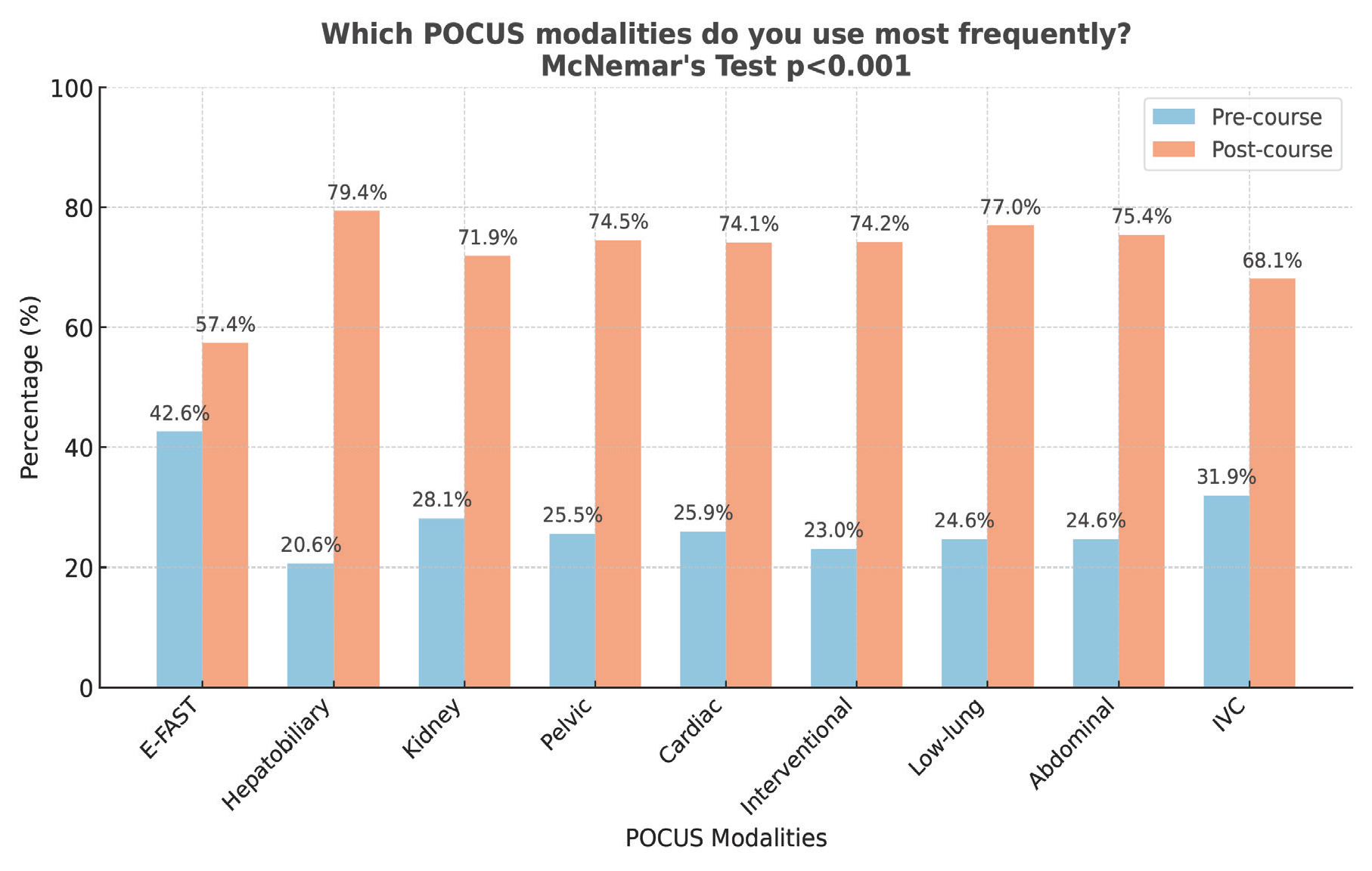
The frequency of POCUS use significantly increased across all scanning modalities post-course (p<0.001). The most prominent increases were noted in hepatobiliary (+58.8%), lower extremity DVT (+54.0%), and abdominal aorta (+50.8%) evaluations ().
POCUS: Point-of-care ultrasound, DVT: Deep vein thrombosis
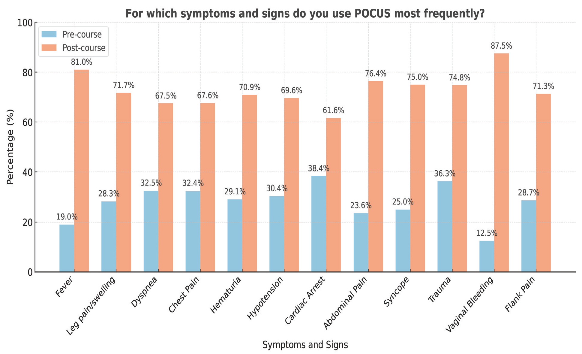
Similarly, POCUS usage increased in relation to various symptoms and signs, most notably in patients presenting with vaginal bleeding (+75.0%), fever (+62.0%), and abdominal pain (+52.8%) ().
POCUS: Point-of-care ultrasound
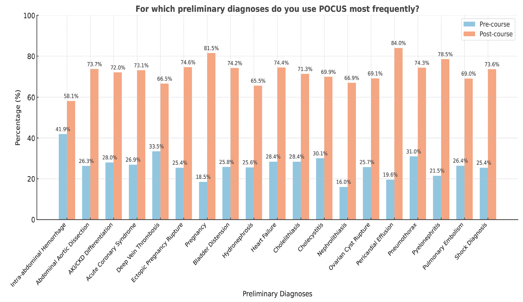
The use of POCUS also rose substantially for preliminary diagnoses, particularly for ovarian cyst rupture (+68.0%), ruptured ectopic pregnancy (+63.0%), and pyelonephritis (+60.8%) ().
POCUS: Point-of-care ultrasound
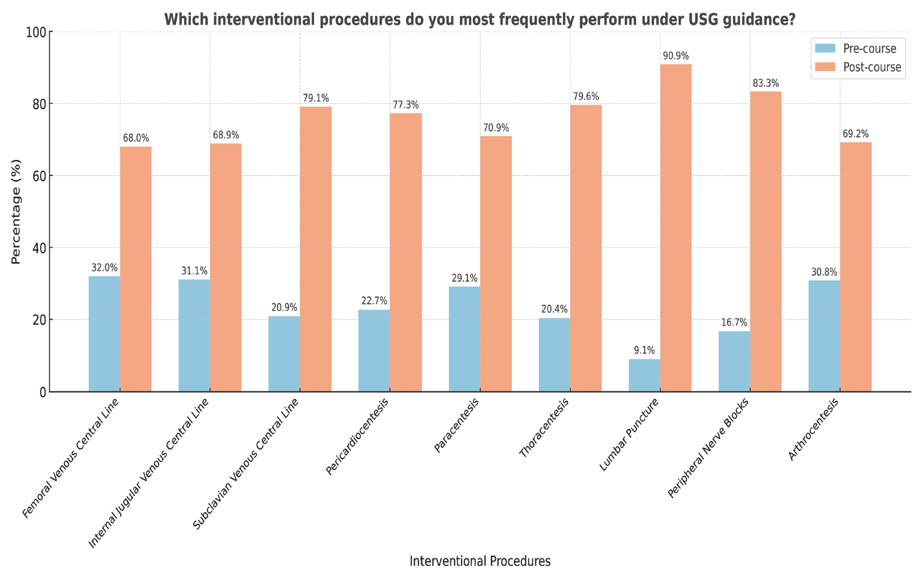
The frequency of POCUS use for interventional procedures increased after the course. The most prominent increases were observed in lumbar puncture (+81.8%), peripheral nerve blocks (+66.6%), and thoracentesis (+59.2%) ().
POCUS: Point-of-care ultrasound,USG: Ultrasonografi
Discussion
The results of this study indicate that participation in a basic POCUS course significantly increased physicians’ use of ultrasound across a wide range of clinical applications. Physicians reported greater diagnostic accuracy and faster decision-making after the training, which aligns with existing literature supporting the integration of POCUS into daily practice ().
Only a subset of physicians responded to the survey (33%). Phillips et al. reported that response rates in survey-based studies in health professions vary widely, often falling below 40% in multicenter studies (-). To minimize non-response bias, we employed standardized electronic invitations and multiple reminders.
The most pronounced increase in usage was observed in hepatobiliary, lower extremity DVT, abdominal aorta, and echocardiographic examinations. These areas reflect both the content emphasized during the course and the clinical relevance of these modalities in emergency care.
Although some physicians were already familiar with E-FAST examinations before attending the course, the relatively modest increase in its post-course use can be attributed to the fact that it is already one of the most widely adopted ultrasound applications among emergency physicians (). Instead, the course seems to have provided a greater boost in confidence and skill in more focused applications, such as DVT, and abdominal aorta scans, which are less commonly used without formal training.
Previous studies similarly demonstrate the impact of structured ultrasound education. Tuvali et al. () showed that even a short POCUS course can have a long-term effect on clinical usage patterns. In a study by Jones et al. (), family physicians reported increased confidence and more frequent use of POCUS in clinical care following training, although their skill retention diminished in areas they did not regularly practice. This supports the view that repeated exposure and clinical reinforcement are essential for sustaining competency.
De Carvalho et al. () and colleagues compared short-term training to longitudinal education and found that longer programs yielded higher frequency of use and greater self-confidence. This suggests that while short courses like the one evaluated in our study are effective for initiating ultrasound practice, longer or repeated sessions may be necessary to preserve skills and deepen clinical integration.
Our findings are also consistent with the results of Rajamani et al. (), where very few participants were able to complete the number of supervised scans required for certification after a short course. Since the basic POCUS course evaluated here did not include a structured follow-up or hands-on assessment, skill retention likely varied among participants depending on clinical opportunities and individual motivation. Implementing supervised scan requirements and certification pathways may improve long-term outcomes and consistency in POCUS practice.
Another key observation is the significant increase in the use of POCUS for specific clinical complaints such as vaginal bleeding, fever, and abdominal pain, as well as for diagnoses like ovarian cyst rupture, ectopic pregnancy, and pyelonephritis. These patterns suggest that the course not only expanded procedural skills but also influenced diagnostic reasoning, promoting earlier use of POCUS in patient workup.
Several studies have addressed the durability of short-term POCUS training, indicating that the gains in knowledge and clinical use can be sustained for weeks to months after the intervention (, ). In our study, the survey was conducted at least six months post-training, supporting the lasting impact of the course. Notably, POCUS use significantly increased for all interventional procedures, particularly lumbar puncture, peripheral nerve blocks, thoracentesis, and pericardiocentesis. Previous research has shown that ultrasound guidance in lumbar puncture improves first-attempt success rates and reduces complications, especially in patients with difficult anatomy (, ). Similarly, ultrasound-guided thoracentesis and nerve blocks enhance procedural accuracy and safety (-). Pericardiocentesis, when performed under echocardiographic guidance, reduces complication rates by optimizing puncture site selection in time-sensitive conditions such as cardiac tamponade (, ). These findings are consistent with our results and highlight how structured training can empower physicians to integrate ultrasound into procedural workflows safely and effectively.
Study Limitations
Although the data show a clear trend toward increased ultrasound use after the course, several methodological limitations should be considered. The survey was completed by only 201 of 610 physicians who were contacted, resulting in a 33% response rate. This raises the possibility of non-response bias, as those more positively influenced by the course may have been more likely to participate. Additionally, participants were asked to retrospectively evaluate their POCUS usage prior to the course, which may have introduced recall bias. The absence of a structured follow-up assessment, supervised practice, or certification component further limits the ability to determine long-term skill retention or clinical competency. Lastly, the sample was composed predominantly of emergency physicians working in tertiary hospitals, which may limit the generalizability of the findings to other specialties and practice settings.
Conclusion
This study demonstrates that even a short, structured POCUS course can lead to a marked increase in physicians’ self-reported use of ultrasound across various diagnostic and procedural domains. While the findings suggest a positive shift in clinical behavior, further research is warranted to evaluate the long-term retention of skills and direct impact on patient outcomes. Broader implementation of standardized ultrasound training, with opportunities for hands-on practice and follow-up assessment, may enhance the integration of POCUS into clinical workflows. Future course designs may benefit from incorporating certification pathways, structured supervised practice, and practical skill evaluations such as objective structured clinical examinations. Additionally, longitudinal training formats, refresher sessions, and re-training opportunities may help reinforce skill retention and optimize long-term clinical integration.
Footnotes
Ethics
Ethics Committee Approval: The study was approved by the University of Health Sciences Türkiye, Fatih Sultan Mehmet Training and Research Hospital Clinical Research Ethics Committee (approval number: 2023/19, date: 12.10.2023).
Informed Consent: Informed consent was obtained from all participants prior to their inclusion in the study.
Acknowledgements
This study was conducted with the permission of the Emergency Medicine Association of Turkey (EMAT). The authors would like to thank the instructors involved in the ultrasound training sessions for their valuable contributions.
Authorship Contributions: Surgical and Medical Practices - B.A.K., M.E., Ö.D.; Concept - B.A.K., M.E., Ö.D.; Design - B.A.K., M.E.; Data Collection or Processing - B.A.K., M.E., Ö.D.; Analysis or Interpretation – B.A.K., M.E.; Literature Search - B.A.K., M.E.; Writing – B.A.K., Ö.D.
Conflict of Interest: No conflict of interest was declared by the authors.
Financial Disclosure: The authors declared that this study received no financial support.
References
Royse CF, Canty DJ, Faris J, Haji DL, Veltman M, Royse A. Core review: physician-performed ultrasound: the time has come for routine use in acute care medicine. Anesth Analg. 2012; 115: 1007-28.
Dietrich CF, Goudie A, Chiorean L, Cui XW, Gilja OH, Dong Y, et al. Point of care ultrasound: a WFUMB position paper. Ultrasound Med Biol. 2017; 43: 49-58.
Atkinson PR, Milne J, Diegelmann L, Lamprecht H, Stander M, Lussier D, et al. Does point-of-care ultrasonography improve clinical outcomes in emergency department patients with undifferentiated hypotension? An international randomized controlled trial from the SHoC-ED investigators. Ann Emerg Med. 2018; 72: 478-89.
Chen Z, Hong Y, Dai J, Xing L. Incorporation of point-of-care ultrasound into morning round is associated with improvement in clinical outcomes in critically ill patients with sepsis. J Clin Anesth. 2018; 48: 62-6.
Abu-Zidan FM. Point-of-care ultrasound in critically ill patients: where do we stand? J Emerg Trauma Shock. 2012; 5:70-1.
Labovitz AJ, Noble VE, Bierig M, Goldstein SA, Jones R, Kort S, et al. Focused cardiac ultrasound in the emergent setting: a consensus statement of the American Society of Echocardiography and American College of Emergency Physicians. J Am Soc Echocardiogr. 2010; 23: 1225-30.
Killu K, Coba V, Mendez M, Reddy S, Adrzejewski T, Huang Y, et al. Model point-of-care ultrasound curriculum in an ıntensive care unit fellowship program and ıts ımpact on patient management. Crit Care Res Pract. 2014; 2014: 934796.
American College of Emergency Physicians. Ultrasound guidelines: emergency, point-of-care, and clinical ultrasound guidelines in medicine. Ann Emerg Med. 2017; 69: e27-e54.
Jang T, Aubin C, Naunheim R. Minimum training for right upper quadrant ultrasonography. Am J Emerg Med. 2004; 22: 439-43.
Levitov A, Frankel HL, Blaivas M, Kirkpatrick AW, Su E, Evans D, et al. Guidelines for the Appropriate Use of Bedside General and Cardiac Ultrasonography in the Evaluation of Critically Ill patients-part II: cardiac ultrasonography. Crit Care Med. 2016; 44: 1206-27.
Gibson LE, White-Dzuro GA, Lindsay PJ, Berg SM, Bittner EA, Chang MG. Ensuring competency in focused cardiac ultrasound: a systematic review of training programs. J Intensive Care. 2020; 8: 93.
Tuvali O, Sadeh R, Kobal S, Yarza S, Golan Y, Fuchs L. The long-term effect of short point of care ultrasound course on physicians' daily practice. PLoS One. 2020; 15: e0242084.
Phillips AW, Reddy S, Durning SJ. Improving response rates and evaluating nonresponse bias in surveys: AMEE Guide No. 102. Med Teach. 2016; 38: 217-28.
Phillips AW, Friedman BT, Durning SJ. How to calculate a survey response rate: best practices. Acad Med. 2017; 92: 269.
Phillips AW, Friedman BT, Utrankar A, Ta AQ, Reddy ST, Durning SJ. Surveys of health professions trainees: prevalence, response rates, and predictive factors to guide researchers. Acad Med. 2017; 92: 222-8.
Ma OJ, Mateer JR, Ogata M, Kefer MP, Wittmann D, Aprahamian C. Prospective analysis of a rapid trauma ultrasound examination performed by emergency physicians. J Trauma. 1995; 38: 879-85.
Jones L, Gathu C, Szkwarko D, Mucheru S, Amin N, Amisi JA, et al. Expanding point-of-care ultrasound training in a low- and middle-ıncome country: experiences from a collaborative short-training workshop in Kenya. Fam Med. 2020; 52: 38-42.
De Carvalho H, Godiveaux N, Javaudin F, Le Bastard Q, Kuczer V, Pes P, et al. Impact of different training methods on daily use of point-of-care ultrasound: survey on 515 physicians. Ultrasound Q. 2024; 40: 46-50.
Rajamani A, Miu M, Huang S, Elbourne-Binns H, Pracher F, Gunawan S, et al. Impact of critical care point-of-care ultrasound short-courses on trainee competence. Crit Care Med. 2019; 47: 782-84.
Henwood PC, Mackenzie DC, Rempell JS, Douglass E, Dukundane D, Liteplo AS, et al. Intensive point-of-care ultrasound training with long-term follow-up in a cohort of Rwandan physicians. Trop Med Int Health. 2016; 21: 1531-8.
Cevik AA, Cakal ED, Abu-Zidan F. Point-of-care Ultrasound training during an emergency medicine clerkship: a prospective study. Cureus. 2019; 11: e6118.
Sanguanwit P, Tansuwannarat P, Bua-Ngam C, Suttabuth S, Atiksawedparit P, Trakulsrichai S. Comparing ultrasonography and surface Landmark-Guided Lumbar puncture in patients with obesity and difficult anatomy; a randomized controlled trial. Arch Acad Emerg Med. 2023; 11: e59.
Mofidi M, Mohammadi M, Saidi H, Kianmehr N, Ghasemi A, Hafezimoghadam P, et al. Ultrasound guided lumbar puncture in emergency department: time saving and less complications. J Res Med Sci. 2013; 18: 303-7.
Boccatonda A, Baldini C, Rampoldi D, Romani G, Corvino A, Cocco G, et al. Ultrasound-Assisted and ultrasound-guided thoracentesis: an educational review. Diagnostics (Basel). 2024; 14: 1124.
Shao RJ, Du MJ, Xie JT. Use of lung ultrasound for the diagnosis and treatment of pleural effusion. Eur Rev Med Pharmacol Sci. 2022; 26: 8771-6.
Haley CB, Beauchesne AR, Fox JC, Nelson AM. Block time: a multispecialty systematic review of efficacy and safety of ultrasound-guided upper extremity nerve blocks. West J Emerg Med. 2023; 24: 774-85.
Alerhand S, Adrian RJ, Long B, Avila J. Pericardial tamponade: a comprehensive emergency medicine and echocardiography review. Am J Emerg Med. 2022; 58: 159-74.
Flint N, Siegel RJ. Echo-guided pericardiocentesis: when and how should ıt be performed? Curr Cardiol Rep. 2020; 22: 71.